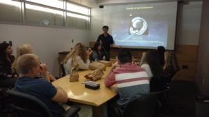
 AP Biology students from Cardinal Wuerl North Catholic High School visited the Department of Structural Biology at the University of Pittsburgh. Students were led on a tour by Dr. Rieko Ishima, an associate professor and a principal investigator in the department. Dr. Ishima oversees a team of research associates and fellows who are currently working to determine protein structure and dynamics using nuclear magnetic resonance.

Protein images are beyond tiny! The nuclear magnetic resonance spectroscopy of proteins does not ‘take a picture.’ Rather, it relies on complex mathematical calculations to build a three dimensional image of the protein.
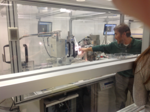 During Dr. Ishima’s tour, students were shown various equipment used in cryo-electron microscopy, nuclear magnetic resonance, and x-ray crystallography. Students were fascinated not only by the incredible detail achieved in the digital images produced by nuclear magnetic resonance (NMR), but also by the sheer size of the equipment required to generate those results.
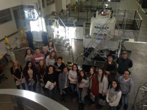 Though NMR examines molecular structure and dynamics at the atomic level, the spectrometers required to view particles that small are extremely large. Pitt has seven spectrometers in this department, and they are housed in 10,000 square foot laboratory. The students were amazed to learn that when the spectrometers were delivered, the first floor windows were removed to allow the equipment to be lowered into the NMR lab! We are standing in front of a two magnets that had to be lowered by crane through an open window.
 Students were also able to tour the cryo-electron microscope facility, where three electron microscopes allow researchers to engage in structural analysis of proteins, viruses, cellular organelles and bacterial cells. Finally, Dr. Ishima and her team led students to the x-ray crystallography lab. Here, researchers are able to grow, store, and monitor crystals. Once crystals are ready for analysis, x-ray beams and image plate detectors are used to collect data about protein structures at the atomic level. While scientists in the lab often use tiny tools to manually transfer crystals for analysis, the lab also is equipped with a robot that can mount and collect data from up to 80 crystals for rapid analysis.
The field is extraordinary.
 On November 18, 2016, AP Biology students from Cardinal Wuerl North Catholic High School participated in a STEM Careers Tour which included a visit to the Department of Developmental Biology at the University of Pittsburgh. Specifically, students were able to interact with Dr. Michael Tsang, an associate professor who is currently conducting research in Pitt’s zebrafish aquaria. In the zebrafish facility, one of the largest in the world, researchers are engaging in multiple large-scale projects which use the zebrafish to understand how organs such as the liver, kidney and heart develop in the embryo.
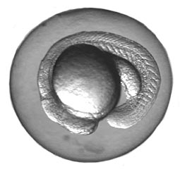 The visit began with a presentation by Dr. Tsang, during which he explained his research and the benefits of experimenting with zebrafish. Students learned that zebrafish are ideal subjects for experimentation because they are small and easily maintained, embryos are transparent and easily visualized during development, and they are able to repair and regenerate damaged tissue. All of the students were fascinated when they learned that, after a few weeks at the bottom of the tank, zebrafish that have sustained a severed spinal cord are able to repair the damage and regain mobility!
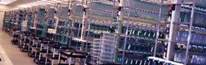 After this presentation, students were able to experience a tour of the zebrafish aquaria, which contains over 11,000 tanks housing over 500,000 zebrafish. While touring the facility, students asked a wide variety of questions about the logistics in place to maintain such a large research lab, and they learned that while the tanks are self-cleaning, university employees spend several hours each day feeding the fish. The rows of tanks with tiny, newly-hatched fish were a highlight of the tour, but the students were most intrigued by the fluorescent green zebrafish. These genetically modified fish carry the gene for Green Fluorescent Protein (GFP), which allows researchers to better identify abnormalities, such as those that lead to Alzheimer’s Disease.
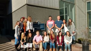 Most importantly, students engaged in dialogue with Dr. Tsang about both the benefits and ethical obligations of animal testing. The visit to the lab, and particularly this conversation with Dr. Tsang, ignited a desire in many of the students to pursue ongoing research with zebrafish. Sixteen AP Biology students from Cardinal Wuerl North Catholic are preparing experimentation results currently being conducted with both adult and embryonic zebrafish for entry into the Pittsburgh Regional Science and Engineering Fair! Mrs. Murray classroom is becoming its own zebrafish aquaria and the contacts she made on the tour have become mentors in her ongoing efforts to make biology come to life for all her students.