 AP Biology students from Cardinal Wuerl North Catholic High School visited the Department of Structural Biology at the University of Pittsburgh. Students were led on a tour by Dr. Rieko Ishima, an associate professor and a principal investigator in the department. Dr. Ishima oversees a team of research associates and fellows who are currently working to determine protein structure and dynamics using nuclear magnetic resonance.

Protein images are beyond tiny! The nuclear magnetic resonance spectroscopy of proteins does not ‘take a picture.’ Rather, it relies on complex mathematical calculations to build a three dimensional image of the protein.
 During Dr. Ishima’s tour, students were shown various equipment used in cryo-electron microscopy, nuclear magnetic resonance, and x-ray crystallography. Students were fascinated not only by the incredible detail achieved in the digital images produced by nuclear magnetic resonance (NMR), but also by the sheer size of the equipment required to generate those results.
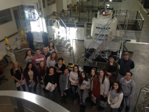 Though NMR examines molecular structure and dynamics at the atomic level, the spectrometers required to view particles that small are extremely large. Pitt has seven spectrometers in this department, and they are housed in 10,000 square foot laboratory. The students were amazed to learn that when the spectrometers were delivered, the first floor windows were removed to allow the equipment to be lowered into the NMR lab! We are standing in front of a two magnets that had to be lowered by crane through an open window.
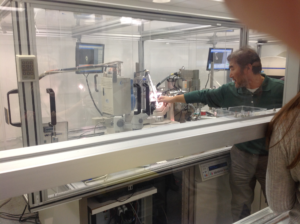
 Students were also able to tour the cryo-electron microscope facility, where three electron microscopes allow researchers to engage in structural analysis of proteins, viruses, cellular organelles and bacterial cells. Finally, Dr. Ishima and her team led students to the x-ray crystallography lab. Here, researchers are able to grow, store, and monitor crystals. Once crystals are ready for analysis, x-ray beams and image plate detectors are used to collect data about protein structures at the atomic level. While scientists in the lab often use tiny tools to manually transfer crystals for analysis, the lab also is equipped with a robot that can mount and collect data from up to 80 crystals for rapid analysis.
The field is extraordinary.